Frank te Nijenhuis
PhD candidate @ ICAI Stroke Lab

I work on applications of AI and computer vision methods in stroke imaging. My main focus is on Digital Subtraction Angiography (DSA), but I also work with other imaging modalities. The goal of my project is to develop tools that can be used to inform decision-making during mechanical thrombectomy.
Within the ICAI Stroke Lab I collaborate closely with my colleagues working on the application of AI methods in the different stages of the (Dutch) stroke care pathway. In addition to the Stroke Lab, I am also a member of the Biomedical Imaging Group Rotterdam, in the Erasmus MC.
I’m supervised by prof. dr. Theo van Walsum, prof. dr. Danny Ruijters, dr. Ruisheng Su and dr. Sandra Cornelissen.
If you are a MSc student looking for a project (either an internship or a full thesis) don’t hesitate to contact me.
Research Interests

Computer Vision for DSA
Developing AI methods for automated analysis of Digital Subtraction Angiography images
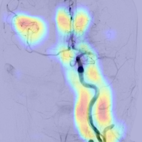
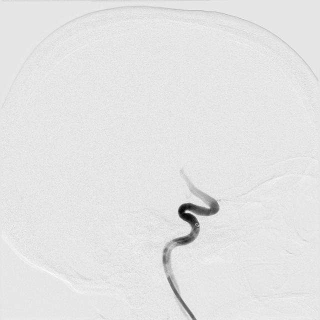
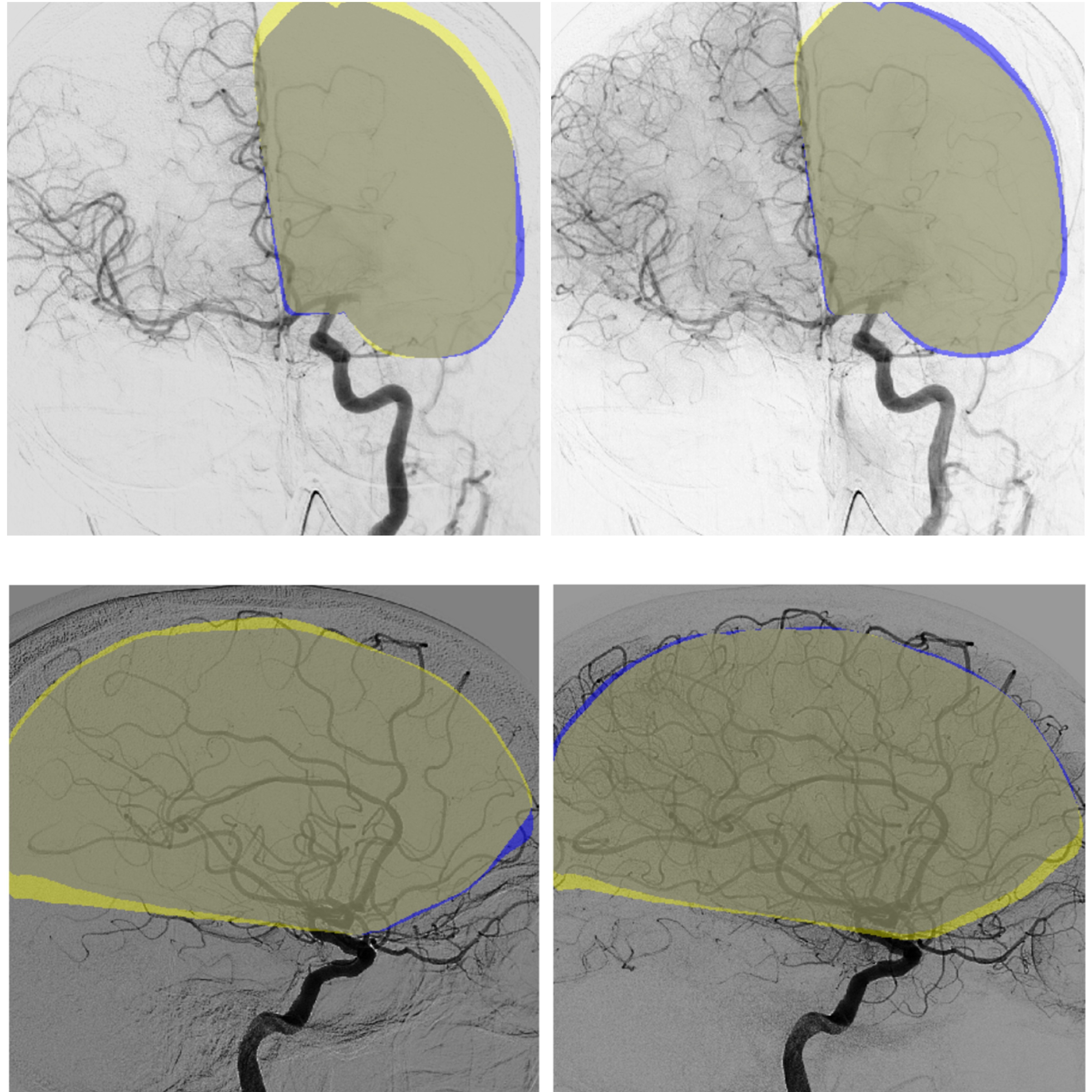
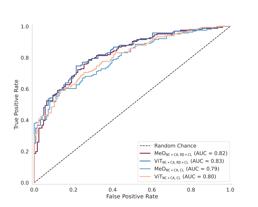
Within my PhD project, we are developing computer vision methods to automatically analyze cerebral Digital Subtraction Angiography (DSA) images. This includes automatic detection of vessel occlusions, quantifying vessel differences between the pre- and post-treatment DSA or delineating brain regions on DSA. The ultimate goal of these methods is to support clinical decision-making during mechanical thrombectomy procedures.

Synthetic Data & Outcome Prediction
Evaluating synthetic tabular data for prediction modelling
With colleagues from the ICAI Stroke Lab we are investigating potential use-cases for synthetic tabular data for prediction modelling. Specifically, we aim to mitigate class imbalance by generating synthetic samples for minority classes, thereby improving predictive performance.

Perfusion DSA
Automatic perfusion imaging in cerebral Digital Subtraction Angiography
Recent work on perfDSA demonstrates the potential of fully automatic and quantitative frameworks for perfusion analysis on DSA, analagous to more established modalities such as CTP and MRP. We are currently validating perfusion DSA to assess its clinical applicability, with the goal of enabling real-time decision-making during thrombectomy.

Carotid Webs
Automatic detection of Carotid Webs on CT Angiography
Carotid webs are shelf-like fibrous projections in the carotid artery that can disrupt blood flow and increase the risk of ischemic stroke. We are developing automated methods to detect these webs on CTA scans, aiming to improve early diagnosis and support clinical decision-making.
Selected Publications
2025
-
 In Stroke Workshop on Imaging and Treatment CHallenges (SWITCH2025) , 2025*These authors contributed equally to this work.
 -
 In Stroke Workshop on Imaging and Treatment CHallenges (SWITCH2025) , 2025
 -
 In Stroke Workshop on Imaging and Treatment CHallenges (SWITCH2025) , 2025


2023
-
 In Stroke Workshop on Imaging and Treatment CHallenges (SWITCH2023) , 2023
